Tuesday, November 13, 2012

GIVE YOURSELF A HAND


The function of the human hand is unlike that of any other animal and its’ anatomy is fascinating and complex.  However, in fitness, the hand is often taken for granted. Understanding hand anatomy and alignment will help you to improve your full body alignment and stability in core conditioning exercises like plank, push ups, and any arm balance poses.

The skeletal structure of the hand & wrist is very similar to the skeletal structure of the feet.  (see previous post: START WITH A STRONG FOUNDATION). There are 27 bones in each hand, which means our hands contain ¼ of the bones in our body.  The bones can be divided into 3 groups: carpals (8 bones), metacarpals (5 bones), and the phalanges (14 bones). 


The muscles of the hand are divided into the intrinsic & extrinsic muscle groups.
Intrinsic muscles are located within the hand itself.  Extrinsic muscles are located in the forearm and attach to the hand through a network of tendons.  The majority of muscles that flex and extend the wrist, and many of the muscles that move the fingers are extrinsic muscles.


The hands are innervated by three nerves: the median nerve (innervates the muscles that help with fine motor or "pinch" function), the ulnar nerve (innervates the muscles that help with a strong hand grasp or grip), and the radial nerve (innervates the wrist extensors helping to control positioning and stability of the hand).

The nerves that innervate the hand all originate in the brachial plexus.  The brachial plexus is a network of nerves that begin at the spinal cord in the neck (cervical spine).  These nerves control movement and sensation in the hand, wrist, elbow and shoulder by carrying messages from the brain to the muscles and other connective tissues. The nerves extend from the brachial plexus and continue down the through the forearm and ultimately enter the hand through small canals in the wrist.  The most well known of these canals is the carpal tunnel.  The median nerve travels through the carpal tunnel to enter the hand.

credit: http://cochiro.com


Just as in the feet, the hand bones form arches that are essential to complex manipulation of objects.  These arches also function as support structures whenever we are bearing weight in our hands.  By maintaining the arches of the hands through proper alignment and use of all of the intrinsic and extrinsic hand muscles, you can keep the nerves free from pressure and injury and create a strong foundation that will translate into better alignment through the entire kinetic chain.


Here is a really great, simple video demonstration of proper hand foundation.  Click on the video "hands foundation".


Next time you practice any exercise that requires bearing weight in your hands, take the extra time to focus on your hand foundation.  You’ll find you can hold plank poses and arm balances longer, increase your push up count, and ultimately improve not only your upper body strength but your core strength as well!


Tuesday, October 2, 2012

IT'S ALL IN THE HIPS


Which muscles first come to your mind when thinking about core stability?  For many people the abdominals are synonymous with the core.  Although the abdominals are certainly part of the puzzle, in order to improve athleticism and prevent injury, many more pieces are needed.

One really great description of the core is a  “ … muscular box with the abdominals in the front, paraspinals and gluteals in the back, the diaphragm as the roof, and the pelvic floor and hip girdle musculature as the bottom” (Akuthota, Ferreiro & Fredericson, 2007).

I covered the abdominals in my last post “Your Abdominals from the Inside Out”.  This post will expand on the core musculature by focusing on the hips and pelvic girdle.  The pelvic girdle holds a position of critical importance when it comes to the kinetic chain.  It is common to see reference to the “lower kinetic chain” (foot, ankle, knee & lumbopelvic hip girdle) and the “upper kinetic chain” (lumbopelvic hip girdle, spine, shoulder, elbow, hand).  The hips and pelvis are the important center link of this chain and play a crucial role in stabilizing the trunk and pelvis in movement and in transfer of force between the upper and lower body.

If you’ve ever watched a professional baseball player, tennis player or golfer you know that it really is all in the hips!  Optimal flexibility and strength in the muscles that support the hips & pelvis coupled with body awareness and endurance is a winning combination for injury prevention and success in any sport.

The bones of the hip joint and pelvic girdle include the three fused bones of the pelvis (ilium, ishcium, pubis), the sacrum (which is actually 5 fused vertebrae at the base of the spine), and the femur.


The joints include the symphysis pubis (where the two pubic bones come together at the front of the pelvis), the two sacroiliac joints (where the sacrum comes together with the ilium at the back of the pelvis), and the hip joint.  The hip joint is a ball and socket joint.  The “ball” is the head of the femur bone.  The “socket” is called the acetabulum and is formed by all three of the pelvic bones (ilium, ischium & pubis).


The first two joints, symphysis pubis & sacroiliac, have very little movement.  The third, the hip joint, allows for movement in a variety of planes.  The muscles that move the hip joint can be divided into the following categories:


Hip Flexors                                                                Hip Extensors
Psoas                                                                           Gluteus maximus
Iliacus                                                                          Hamstrings
Rectus Femoris                                                          
Sartorius
Muscles of the medial compartment of the thigh (pectinius, adductor longus, adductor brevis, gracilis)

Adductors                                                                   Abductors
Adductor brevis                                                          Gluteus medius
Adductor longus                                                         Gluteus minimus
Adductor magnus & minimus
Pectineus
Gracilis
Obturator externus

Lateral Rotators                                                        Medial Rotators
Obturators (internus & externus)                                Gluteus medius
Gemelli (superior & inferior)                                      Gluteus minimus
Piriformis
Quadratus femoris

___________________________________________________________


HIP FLEXORS


________________________________________________________


GLUTEUS MAXIMUS 


GLUTEUS MEDIUS


 __________________________________________________________

LATERAL ROTATORS (credit: www.iadms.org)
_____________________________________________________

ADDUCTORS


____________________________________________________

Due to the central location of the hips and pelvis in the kinetic chain, imbalances in the strength and flexibility of the hip muscles can result in misalignment and injury farther up and down the kinetic chain.

A great example of this is the effect of weak gluteal muscles on the knee and foot.  If gluteus medius and gluteus minimus (our primary hip abductors) are weak, the femur will tend to adduct and internally rotate.  If you follow this down the kinetic chain, the knee will fall into a “knock-kneed” position and proper knee tracking will be disturbed, the foot will tend to pronate.  The smaller leg muscles are not able to make up for the weakness of the gluteal muscles and a number of injuries (IT band syndrome, achilles tendionosis, plantar fasciitis, & shin splints) can result along the lower kinetic chain. 

Since over activity of the adductor muscles coupled with weakness of the gluteal muscles is one of the most common imbalances that can lead to injury lets look at a couple of exercises you can easily add to your fitness routine to help prevent this imbalance.

The gluteal muscles are primarily hip extensors and abductors.  Exercises that involve extending your leg behind you and lifting your leg to the side will target these muscles.  The tricky part, especially in people who have trouble accessing these muscles, is making sure the gluteal muscles are doing the work.  Your abdominals are the key to keeping your pelvis and spine stable during hip extension work.

Here is a nice pictorial of the “Bird-Dog” or “all fours” exercise from ACE fitness http://www.acefitness.org/exerciselibrary/14/bird-dog
Although it looks quite simple, I would suggest starting this exercise lifting your leg only.  Then progress by taking your opposite hand on to your abdominals while you lift your leg.  This is a great way to provide some sensory feedback as to the stability of your spine and pelvis.  Your “hip-bones” (the bones you feel protruding on the front of your pelvis) should stay in the same vertical plane as your pubic bone to maintain neutral pelvis, and you should feel your abdominals pulling in toward your spine.

You can then add abduction by maintain the height of your leg and moving it away from the midline of your body.  Again, try this with your opposite hand on your abdominals to help police the stability of your spine and pelvis as well as the depth of your abdominal contraction.

Lunges are a fantastic exercise for simultaneously strengthening and stretching the hip adductors.  Here is a video link demonstrating a very basic lunge. http://www.ehow.com/video_2365368_do-basic-lunge-leg-exercise.html


Training all of the muscles of the hip in a way that balances strength and flexibility will not only prevent local injury but will also help to maintain alignment and prevent injury along the whole length of the kinetic chain.


Akuthota, V.A., Ferreiro, T.M. Fredericson, M. (2007) Core stability exercise principles. Current Sports Medicine Reports, 7(1), 39-44.

Geraci M.C. (1994) Rehabilitation of pelvis, hip and thigh injuries in sports. Physical Medicine & Rehabilitation Clinics of North America, 5, 157-73.

Geraci, M.C., Brown, W. (2005) Evidence-Based treatment of hip and pelvic injuries in runners. Physical Medicine & Rehabilitation Clinics of North America, 16, 711-747.

Lloyd-Smith, R., Clement, D.B., McKenzie, D.C., et.al. (1995) A survey of overuse and traumatic hip and pelvis injuries in athletes. The Physician and Sportsmedicine, 13, 131-41.

Sciascia, A., Cromwell, R. (2012) Kinetic chain rehabilitation: a theoretical framework. Rehabilitation Research and Practice, 2012, 1-9.


Tuesday, August 28, 2012

Your abdominals from the inside out


Your mom probably told you long ago that it’s what’s on the inside that counts.  Nothing could be closer to the truth when it comes to training your abdominals.  That may sound a bit strange considering that most people looking to firm and sculpt their abdominals spend hours each day crunching away at the gym with visions of tight, flat, tummies dancing in their heads.  To understand how to effectively train your abdominals for optimum spinal health as well as to get the look you desire, we’ve got to explore the abdominal anatomy from the inside out.

There are four layers of abdominal muscles:
  • Transverse abdominis
  • Internal obliques
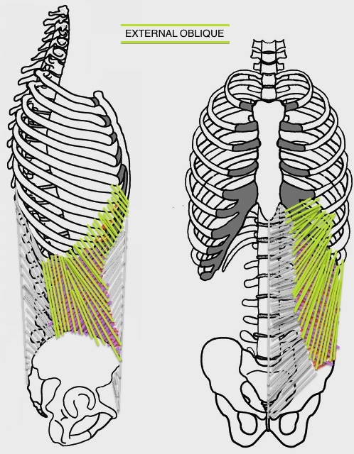
  •  External obliques
  • Rectus abdominis


Transverse Abdominis is the deepest layer of abdominal muscle.  It functions as your body’s corset, supporting the contents of the abdominal cavity and providing spinal stability.  This muscle originates on the iliac crest (top of the pelvis), the lower six ribs, and the sheath of connective tissue that covers the deep muscles of the back and trunk (thoracolumbar fascia).


The transverse abdominis is a muscle of stability, not of movement.  Beginning in the late 1980’s, the use of imaging techniques to directly measure the activity of this muscle has allowed more insight into the role of transverse abdominis in spinal stabilization. 

Transverse abdominis activates subconsciously in anticipation of trunk and limb movement.  Unlike the other abdominal layers, transverse abdominis is recruited continuously regardless of the direction of movement.  Contraction of this muscle reduces the circumference of the abdominals (flattening you from the front and slimming you from the sides).  This circumferential contraction (with the aid of the pelvic floor and diaphragm muscles) creates an air-bag effect providing support and stability to your spine.  Studies have shown that, in people with back pain, contraction of transverse abdominis is often delayed, disturbed, and sometimes completely absent.

How to access and train your transverse abdominis:


To find your transverse abdominis, hook your fingers on the bony protuberances on the front of your pelvis (your “hip bones”) and imagine pulling a drawstring tight from hip bone to hip bone … you should feel this muscle engage laterally across the front of your pelvis.

Once you've located the muscle, dead bug is a great exercise to help you isolate and strengthen it. 

Begin lying flat on your back, knees bent, feet hip distance apart and spine and pelvis in neutral. (In neutral spine you maintain the natural curvatures of your spine.  In neutral pelvis your “hip bones” should be in the same vertical plane as your pubic bone).  Engage your pelvic floor muscles, engage transverse abdominis, and bring your knees over your hips while reaching your arms for the ceiling.  Slowly move one leg forward while reaching the opposite arm back.  You will use your transverse abdominis to prevent any movement in the spine or pelvis.  If this proves too challenging, you can place your arms and/or one foot on the floor.

If you are having trouble feeling the contraction of transverse abdominis, give yourself as much sensory feedback as possible.  Place a hand on your abdominals to provide tactile feedback and/or perform the exercise in front of a mirror to give yourself visual feedback as to the stillness of your spine and pelvis. 

CLICK HERE for  a nice video link of a physical therapist demonstrating the dead bug exercise.

The remaining layers of abdominal muscles are involved in movement of the trunk.  Your trunk & spine can move in a multitude of directions including flexion, extension, lateral bending and rotation. The deep and superficial back muscles work with the abdominal muscles to create trunk movement.


TRUNK MOVEMENT
MUSCLES USED
Flexion
Rectus abdominis, internal & external obliques, psoas
Extension
Erector spinae, quadratus lumborum, lower trapezius, interspinalis
Lateral bending
Quadratus lumborum, internal and external obliques, transverse abdominis, intertransverse
Rotation
Internal & external obliques, erector spinae, quadratus lumborum, transverse abdominis, multifidus, rotatores, psoas


To keep things simple, you can take the same dead bug exercise described above and layer on work for your oblique muscles and rectus abdominis.  Be sure that you've mastered the basic exercise described earlier before adding on these variations.

From the same starting position (knees over hips, arms reaching for the ceiling), use your oblique muscles to create a posterior pelvic tilt (lifting the front of your pelvis toward your low ribs, pressing your lower back in to the floor).  Now challenge the contraction in the same way you did in the first exercise (opposite leg and arm reaching away from each other). You may find you have greater range of motion in the exercise now that you’ve added another layer of abdominal contraction.

Finally, add a challenge for the outer layer of abdominals by straightening your legs over your hips, then, keeping your low back on the floor slowly reach both legs away from you (this provides an eccentric contraction for the outer layer of abdominal muscle – lengthening the muscles while they contract).  Bring your legs back in over your heart and roll your tailbone and sacrum up off the floor (be sure to keep your shoulders firmly on the floor – use only your abdominals!). 

Remember to start from the inside out in any abdominal exercise that you choose to perform.  Find stability from your deepest layer of abdominal muscles and then add movement to train the outer layers.  The result will be the winning combination of firm, flat abdominals & a happy, healthy spine!

References: